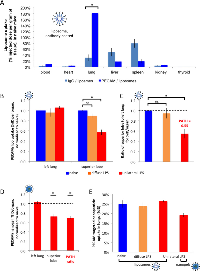
Figure 2. PECAM/NCs markedly concentrate in the lungs, but preferentially accumulate in healthy rather than inflamed lung tissue.
(A) (A) Healthy (naïve) mice were IV-injected with liposomes (inset; labeled with I125) conjugated to anti-PECAM antibodies or control antibodies, sacrificed 30 minutes later, and organ uptake was measured by gamma counter. Anti-PECAM liposomes display 5.9-fold higher lung concentratinos than control liposomes. (B) 24 hours after either unilateral LPS (red bars), diffuse LPS (orange), or sham (naive; blue), mice were IV-injected with anti-PECAM-liposomes, sacrificed 30 minutes later, and then lung lobes were measured as in A. For each mouse, we calculated the ratio of superior lobe to left lung for the percent injected dose (%ID) per organ (“organ” here being a lung lobe), and normalized to naive. (C) For each mouse, we calculated the ratio of superior lobe to left lung for the %ID/organ values in B, normalized to the value for naïve mice. Note that this ratio for unilateral LPS mice (third bar) is defined as the PATH ratio (Pathologically Altered To Healthy ratio), which is the %ID/organ of the pathological (inflamed) tissue divided by the %ID/organ of the healthy tissue, normalized to naive. A PATH ratio < 1 (here it is 0.55), indicates that a NC preferentially accumulates in healthy tissue, while a PATH ratio > 1 implies preferential accumulation in pathological (inflamed) tissue. (D) With the same protocol as in B and C, unilateral LPS mice were injected with an alternative nanocarrier, nanogels, also coated with anti-PECAM antibodies, producing a similar PATH ratio < 1. Only values with unilateral LPS are displayed (red bars). (F) Replotting A-D as %ID shows no differences in whole-lung uptake of PECAM/NCs, regardless of the presence of LPS instillation. *, p < 0.05. ns, non-significant.

