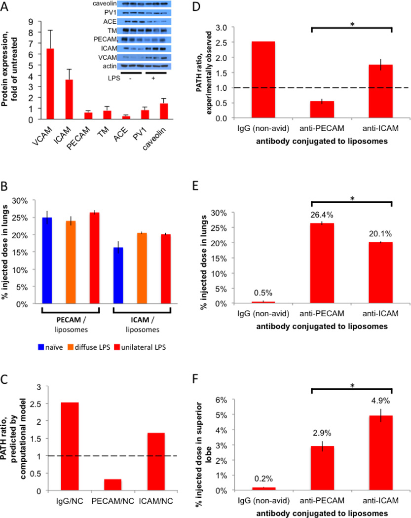
Figure 6. ICAM/NCs accumulate preferentially in inflamed lung regions because of increased epitope levels in that region.
(A) A screen to identify targeting epitopes that are increased in the lungs of mice after LPS stimulation. Inset, Western blot of whole lung homogenates from either naive mice or mice given IV LPS 24 hours before sacrifice. Each lane represents an individual mouse, 3 mice per condition. The bar graph shows quantification of the inset Western, normalized to actin, and then mean of LPS mice was divided by the mean of naive mice. (B) ICAM/liposomes display increased (1.26x) whole-lung uptake in diffusely inflamed lungs, but still have lower (by 15%) whole-lung uptake than PECAM/liposomes. 24 hours after either unilateral LPS (red bars), diffuse LPS (orange), or sham (naive; blue), mice were IV-injected with I125-labeled liposomes and whole-lung uptake measured 30 minutes later. (C) A multi-compartment pharmacokinetic model (see Supplemental Materials), incorporating our measurements of hypoxic vasoconstriction, capillary leak, and epitope expression, predicts a PATH ratio < 1 for PECAM/NCs (first red bar) but a PATH ratio ∼1 for ICAM/NCs. (D) Experimentally determined PATH ratios show that PECAM/NCs preferentially accumulate in healthy lung tissue (PATH ratio < 1), while ICAM/NCs are preferentially taken up into the inflamed superior lobe (PATH ratio > 1). (E) Whole-lung uptake, shows PECAM/NCs have the greatest total lung uptake. (F) Uptake in the just the intended target lobe, the severely inflamed superior lobe, with ICAM/NCs performing the best. *, p < 0.05.

