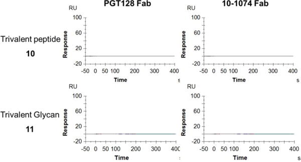
Figure 3.

SPR sensorgrams of the binding between antibody Fab fragments and nonglycosylated trivalent V3 peptide or trivalent glycans. a) with trivalent V3 peptide (10); b) with trivalent Man9GlcNAc2 glycan (11). Fabs were run from 1000 nM with 1:2 serial dilutions. Data were fit with a 1:1 Langmuir binding model (fitting was shown in black). Ka was given in M−1S−1; Kd in S−1 and KD in M.S1)
