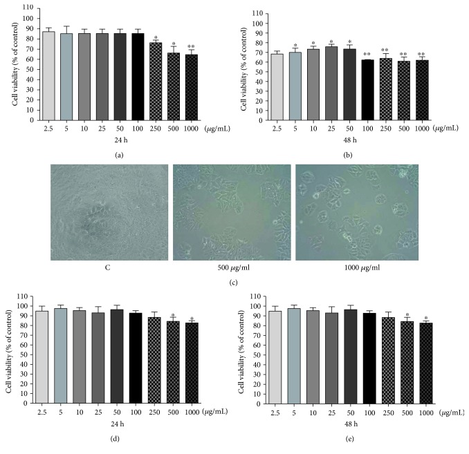
Figure 1.
Effect of PE (2.5–1000 μg/mL) on viability of MCF-7 (a, b) and MDA-MB-435 (d, e) cells at different time intervals after exposure using MTT assays. The experiment is expressed as mean ± standard error, and differences significant between treated cells with PE were compared using the Tukey test (∗p < 0.05; ∗∗p < 0.01). Phase contrast microscopy of MCF-7 cells (treated for 48 h with 500 and 1000 μg/mL of PE) was observed on 96-well culture plates (c).

