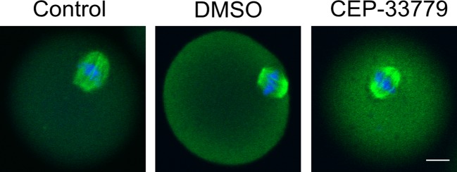
Figure 3. CEP-33779 does not disorder microtubules reorganization in mouse oocytes.
Metaphase I(M I) spindle morphology of mouse oocytes were observed during meiosis in control (n=77), DMSO (n=83), and CEP-33779 (n=72) groups. DNA (blue) and tubulin (green) were stained with DAPI and antitubulin antibody, respectively. The scale bar is 20 μm.

