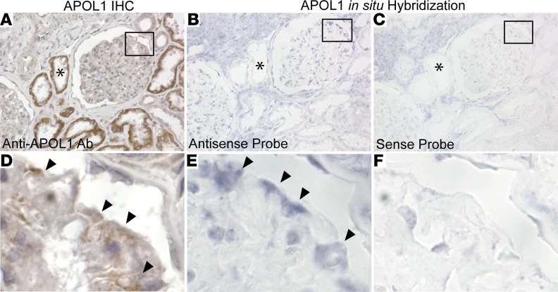
Figure 1. Apolipoprotein L1 mRNA in a normal human kidney by in situ hybridization.
(A–C) Representative images from normal human kidney tissue obtained from two tumor nephrectomies. Serial sections showing apolipoprotein L1 (APOL1) expression by immunohistochemistry (A) (color reaction product is brown, counterstained with hematoxylin) and with in situ hybridization using antisense probe (B) (color reaction product is blue, with no counterstain) and sense probe as control (C). APOL1 protein and mRNA expression is present in the glomerulus but in contrast to the strong positive signal (denoted by asterisks) detected by immunohistochemistry (A); a weak in situ hybridization signal is seen in some but not all proximal tubules (B). (D–F) Magnification of boxed regions in A–C shows that a positive signal is present in podocytes (arrowheads) for both staining methods. Original magnification, ×40 (A–C); ×285 (D–F).

