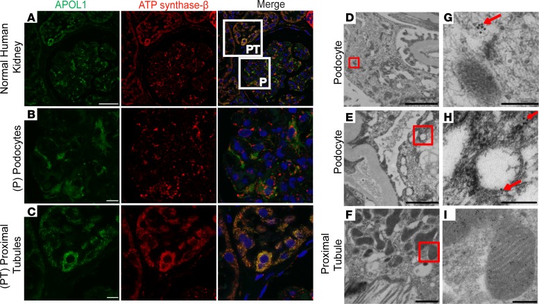
Figure 2. Subcellular localization of apolipoprotein L1 in normal human kidney.
(A–C) Confocal immunofluorescence microscopy images showing apolipoprotein L1 (APOL1) (green) colocalizing with the β subunit of ATP synthase (red) in proximal tubules of normal human kidneys (C) but not in podocytes (B). (B and C) Magnified images of the boxed regions shown in the merged image of A, showing the podocyte (B) and the proximal tubule (C). Nuclei were stained with TOTO-3 iodide (scale bars: 10 μm). (D–F) Immunogold electron microscopy image of human podocyte and proximal tubules from a normal human kidney (scale bars: 2 μm). (G–I) Magnification of the boxed regions from D–F demonstrates (G) gold particles in cytoplasm associated with podocyte cytoskeleton (red arrow), (H) vesicles in podocytes (red arrows), and (I) gold particles in proximal tubules localizing to mitochondria (scale bars: 0.2 μm).

