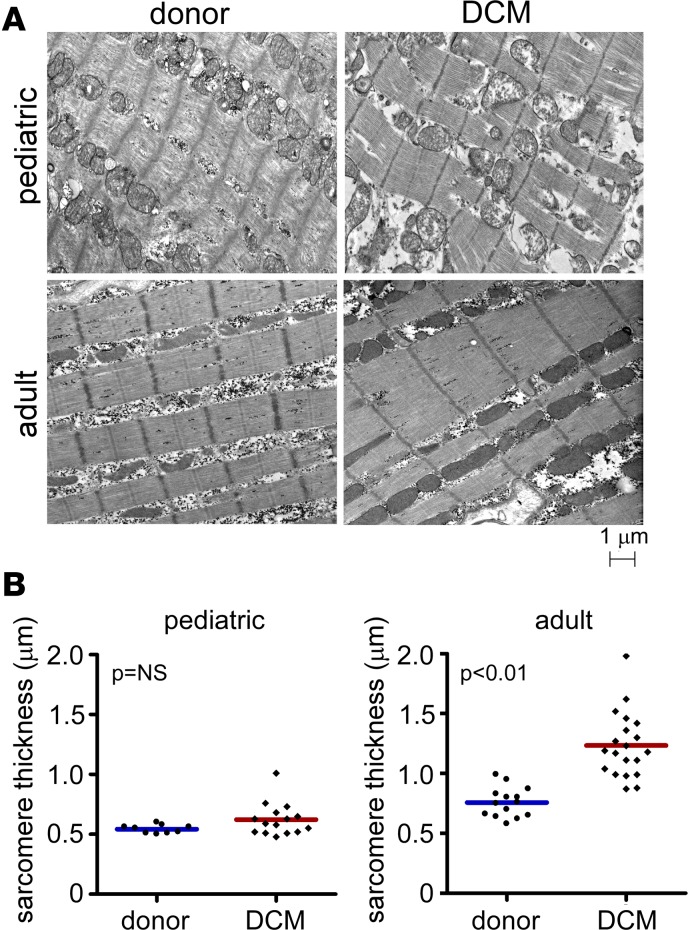
Figure 2. Sarcomere structure in pediatric and adult DCM.
(A) Electron microscopy examining sarcomere structure in donor control, pediatric, and adult DCM specimens. Compared with donor controls, pediatric DCM patients display no change in sarcomere thickness. In contrast, adult DCM patients demonstrate increased sarcomere thickness compared with donor controls. (B) Quantification of sarcomere thickness. Each data point represents an average of >20 sarcomeres measured from individual patient samples. P values found with Mann Whitney U test.

