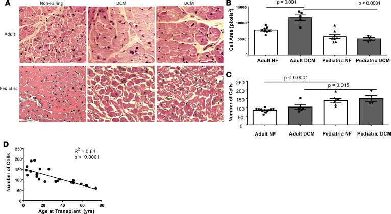
Figure 8. Pediatric and adult DCM cell area and number from 4 dilated cardiomyopathy (DCM) and 7 nonfailing (NF) pediatric hearts and from 4 DCM and 9 NF adult hearts.
(A) Representative images from adult and pediatric NF and DCM left ventricle (LV). 40× magnification. (B) Cardiomyocyte area quantified from adult and pediatric NF and DCM LV. Cardiomyocytes from adult DCM LV have significantly increased area when compared with cardiomyocytes from adult NF LV and pediatric DCM LV (adult DCM vs. adult NF, P = 0.001; adult DCM vs. pediatric DCM, P < 0.0001 [ANOVA with Sidak’s correction]). (C) Quantification of the number of myocytes in adult and pediatric NF and DCM LV. There is a significantly greater number of myocytes in pediatric NF LV compared with adult NF LV. P < 0.0001 (ANOVA with Sidak’s correction). (D) The number of cardiomyocytes correlates with the age of the patient at the time of transplant (or donation) as determined by Spearman rank correlation.

