
Figure 3.
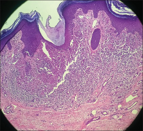
Section shows hyperkeratosis and irregular epidermal acanthosis with saw-tooth rete pegs and focal basal layer vacuolar degeneration. The superficial dermis and dermoepidermal junction show lymphohistiocytes in a band-like pattern suggestive of lichen planus hypertrophicus. (H and E, ×200)