Figure 4.

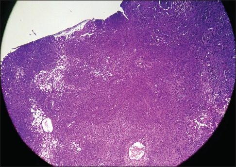
Sections show sheets of proliferating keratinocytes and tiny squamous pearls with mild pleomorphism of keratinocytes suggestive of well-differentiated squamous cell carcinoma. (H and E, ×400)

Sections show sheets of proliferating keratinocytes and tiny squamous pearls with mild pleomorphism of keratinocytes suggestive of well-differentiated squamous cell carcinoma. (H and E, ×400)