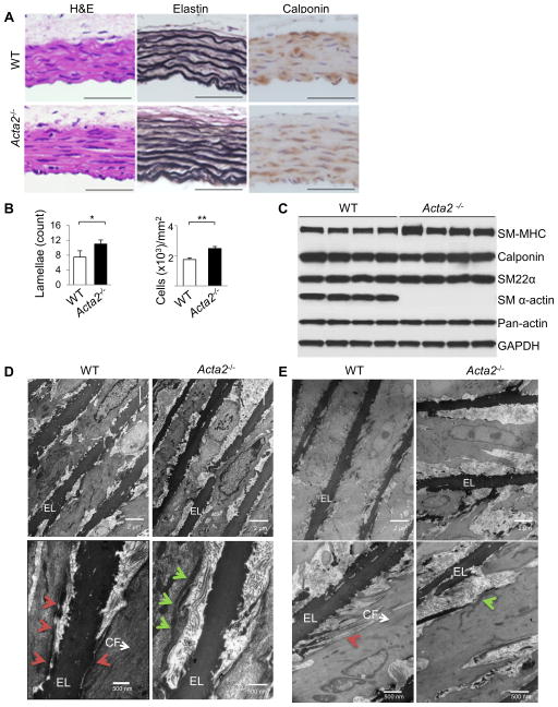
Figure 1. Ultrastructural defects in Acta2−/− mouse aortas.
(A,B) Histology from WT and Acta2−/− aortas at 4 weeks of age shows increased elastin layers and increased density of SMCs marked by calponin staining (n=3 per genotype, *p<0.05, **p<0.01). (C) Western blot analysis of 4 individual WT and Acta2−/− mice shows comparable levels of aortic SMC contractile protein expression at 4 weeks of age. SM α-actin is absent in Acta2−/− aortas as expected, but another actin isoform is expressed to compensate based on similar pan-actin levels for both genotypes. (D,E) Electron microscopy analysis of Acta2−/− aortas at 4 weeks (D) and 6 months (E) of age shows a lack of contractile filaments (CF) within SMCs, aberrant connections between the cells and the elastin fibers (EL), and submembranous dense plaques (green arrows) rather than focal adhesions (red arrows) (n=2 mice per genotype per timepoint).

