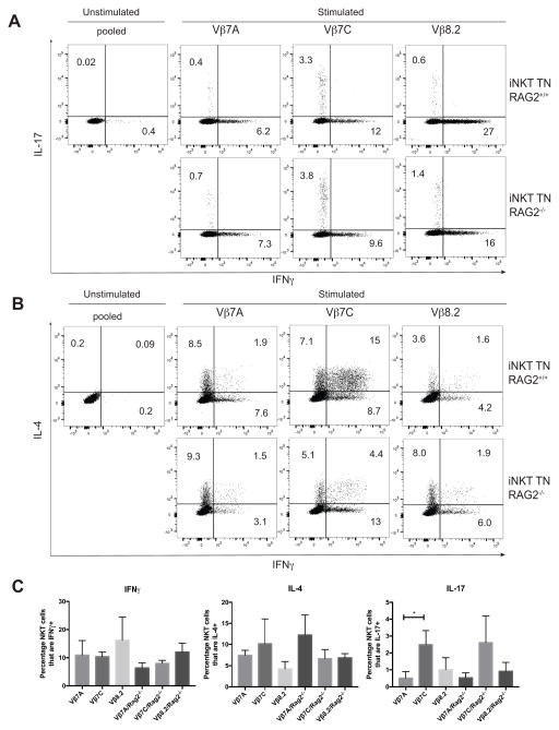
Figure 5. NKT1, NKT2 and NKT17 subsets are present in rigorously monoclonal mice.
(A–B) Skin draining lymph node cells from all TN mice were stimulated in vitro with PMA/Ionomycin for 4 hours, in the presence of GolgiStop. Cells were stained with anti-CD3 and CD1d (PBS57) tetramer, before being fixed, permeabilized, and stained with antibodies to IFNγ, IL-4, and IL-17. Results shown are gated on CD3+CD1d-tetramer+ cells. TN mice are either RAG-proficient (top panels) or RAG-deficient (bottom panels). (C) Quantification of panels A–B, n=4 mice per RAG2+/+ group and n=3 mice per RAG2−/− group. Mice were 5–6 weeks of age and both sexes were included in each group.

