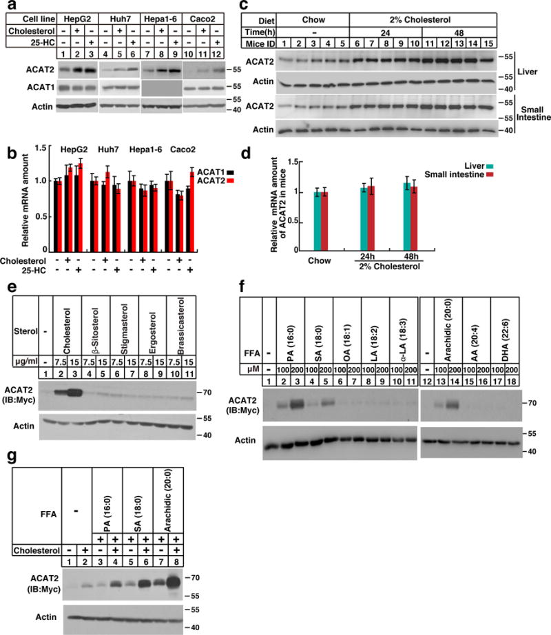
Figure 1. ACAT2 is stabilized by sterols and saturated fatty acids.

(a–b) Cells (HepG2, Huh7, Hepa1-6 and Caco2) were depleted of lipids by incubating in medium supplemented with 5% LPDS, 1 μM lovastatin, 50 μM mevalonate for 16 hrs. Then the cells were treated without (−) or with (+) sterols (15 μg/mL cholesterol and 3 μg/mL 25-HC) for 16 hrs. The cells were harvested for western blotting and RT-qPCR (n=3 independent experiments, mean±S.D.).
(c–d) Male C57/BL6 mice (8–12 weeks) were fed on chow diet or high cholesterol diet (HCD, chow diet supplemented with 2% cholesterol) for 24 and 48 hrs. The liver and small intestine samples were subjected to western blotting and RT-qPCR analysis (mean ± S.D, n=5 mice).
(e–g) The CHO/ACAT2-Myc cells were depleted of lipids as in Figure 1a. Then the cells were treated with different sterols (e), fatty acids (f) and cholesterol together with FAs (g) at indicated concentration. After incubation for 16 hrs, the cells were harvested for western blotting.
The immunoblots are representative of at least 3 independent experiments. Uncropped blots are shown in Supplementary Fig. 8. Statistics source data for b, d can be found in Supplementary Table 2.
