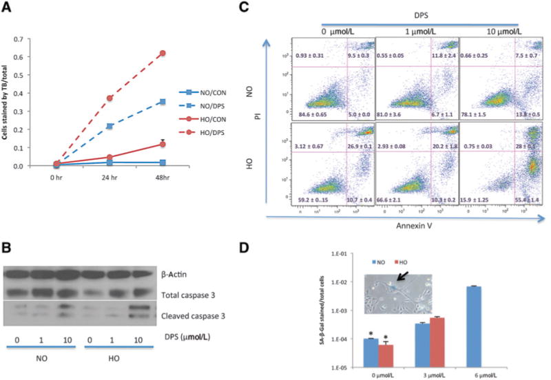
Figure 4.

Darinaparsin induced apoptosis and senescence in HI-LAPC-4 cells. A, cells were treated with darinaparsin (10mmol/L) for the time indicated and analyzed with Trypan blue exclusion assay. B and C, cells were treated with darinaparsin (24 hours) under NO and HO before measurements of cleaved caspase-3 (B, Western blot) and Annexin V externalization (C, flow cytometry). D, cells were treated with darinaparsin (24 hours) under NO or HO, and maintained in fresh medium for 24 hours before SA-β-Gal staining. Few cells survived the darinaparsin treatment (6 μmol/L) under HO, and therefore were not counted. P< 0.001 by 1-wayANOVA, and *,P< 0.001 versus 3 and 6 μmol/LbyTukey multiple comparison test. The arrow of the inset indicates a cell with senescence morphology and positive stain of SA-β-Gal.
