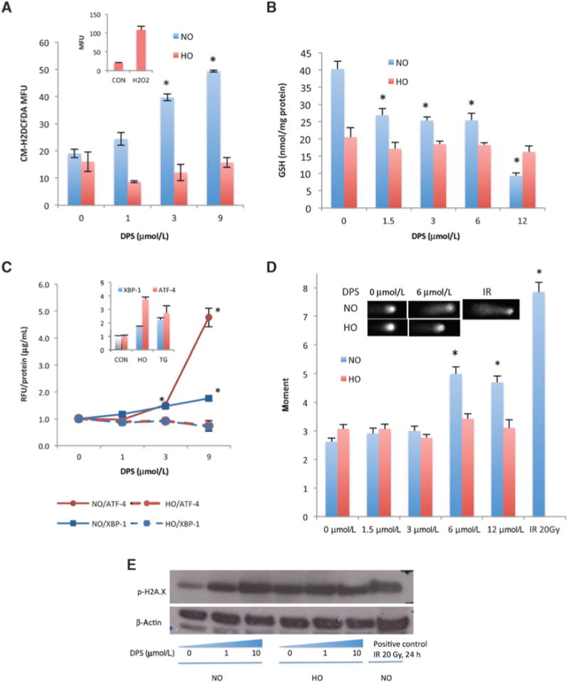
Figure 5.

Darinaparsin induced oxidative stress under normoxia but not hypoxia. A, HI-LAPC-4 cells were treated with darinaparsin (8 hours) and assayed with fluorescent probe CM-H2DCFDA. Inset, ROS level of cells with or without H2O2 (positive control) treatment (1 mmol/L, 10 minutes) under HO. B, HI-LAPC-4 cells were treated with darinaparsin (4 hours) before analysis for the total cellular GSH content. C, pancreatic cancer (MIA PaCa-2) cells that stably express a luciferase reporter of ATF-4 or XBP-1 were treated with darinaparsin (8 hours) and thapsigargin (TG, 30 nmol/L, 24 hours, positive control). The luciferin production was expressed as the relative fluorescence intensity (RFU) over untreated control, and normalized by protein concentration. Inset: both HO and TG induced XBP-1 and ATF-4 activity. D and E, HI-LAPC-4 cells were treated with darinaparsin for 4 hours and used for comet assay (D) and γH2AX Western blot (E).IR, irradiation (positive control). Inset, representative comet micrograph. P< 0.0001 for 1-way ANOVA in A-E comparing conditions under NO. *,P< 0.01 for comparison with NO untreated control by the Tukey multiple comparison test.
