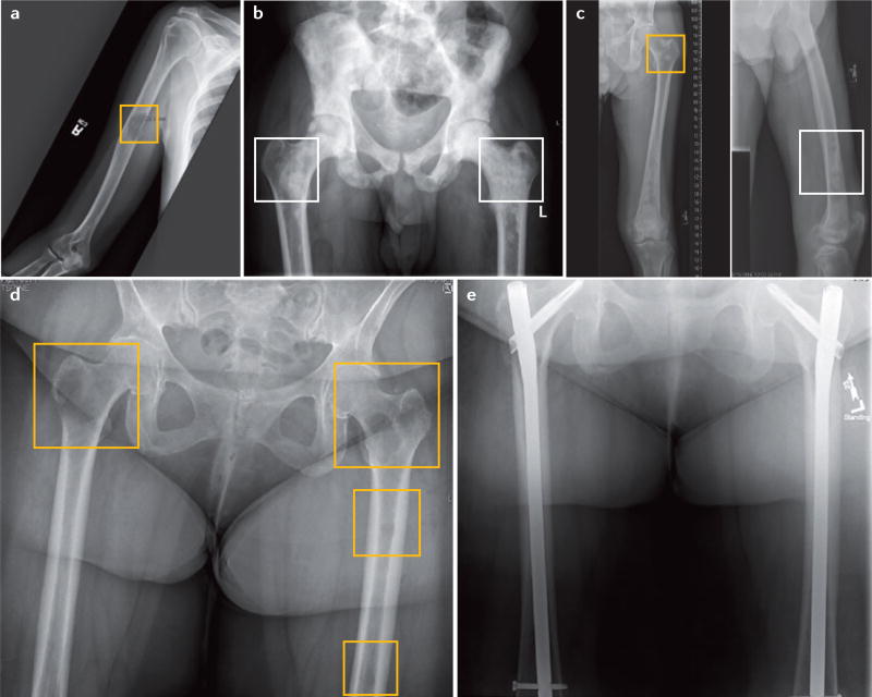
Figure 2. Presentation of lytic, blastic and mixed bone lesions.
a | Anterior–posterior radiograph of the humerus demonstrating a mid-shaft lytic lesion (yellow square), in which >50% of the cortical bone has been destroyed. The radiograph is consistent with osseous involvement by metastatic adenocarcinoma of the lung. b | Anterior–posterior radiograph of the pelvis demonstrating diffuse blastic lesions involving the pelvis and bilateral proximal femurs (white squares). The radiograph is consistent with diffuse osseous involvement by metastatic prostate cancer. c | Anterior–posterior radiograph of femur demonstrating mixed blastic (white square) and lytic lesions (yellow square) involving the proximal femur and shaft from a patient with hepatocellular carcinoma. d | Anterior–posterior radiograph of bilateral femurs demonstrating multiple lytic lesions in both femurs (yellow squares). Both femurs are at considerable risk of fracture. The radiograph is consistent with osseous involvement by metastatic breast cancer. e | Postoperative anterior–posterior radiograph of bilateral femurs (from part d) after radiotherapy and surgical treatment with intramedullary nails for prophylactic stabilization of impending fractures and improved patient quality of life.

