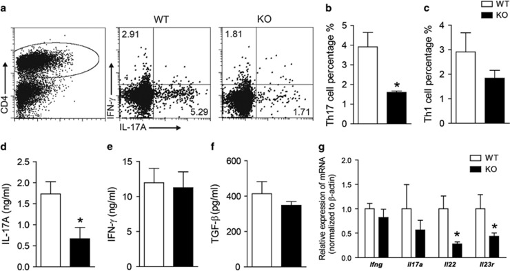
Figure 3.
Gαq-KO inhibits in vivo Th17 development. Leukocytes were isolated from the lymph nodes of WT mice and Gαq-KO mice on day 10 PI and analyzed with flow cytometry. (a–c) Th1 and Th17 cells were analyzed by intracellular staining of IFN-γ and IL-17A, respectively, in the CD4+ gate. (d–f) Lymph node leukocytes from EAE-induced WT mice and Gαq-KO mice were re-stimulated in vitro with MOG35–55 (20 μg/ml) for 72 h, and IL-17A (d), IFN-γ (e) and TGF-β (f) in supernatants were analyzed with ELISA. (g) qPCR analysis of Th1- and Th17-related gene expression in lymph nodes. The data are expressed as the mean±s.e.m. (n=3), * P<0.05 versus WT-EAE.

