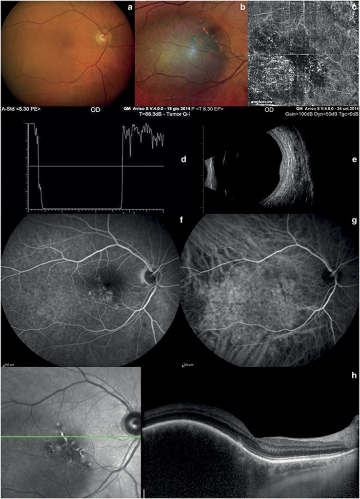
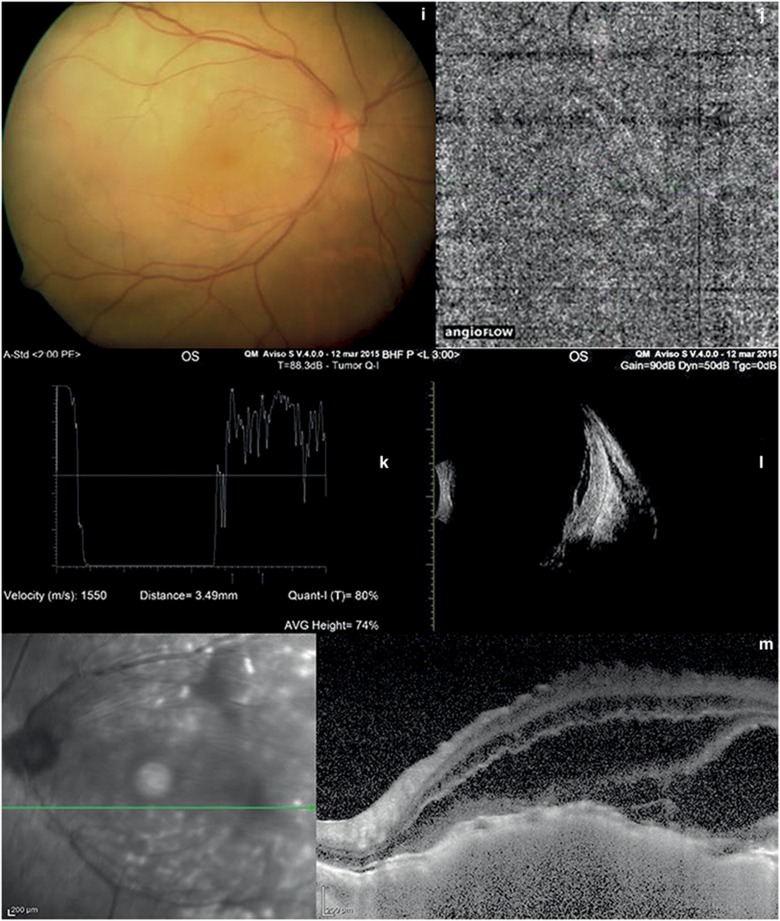
Figure 3.
Choroidal hemangioma and choroidal metastasis. (a and b) Color fundus photography of hemangioma and multicolor imaging show an orange–red lesion. (c) OCT-angiography of hemangioma shows large choroidal vessels inside the tumor. (d) A-scan echography of hemangioma shows high reflectivity of the lesion. (e) B-scan echography of hemangioma shows a dome-shaped hyperreflective mass. (f, and g) Fluorescein and indocyanine angiography of hemangioma reveal hyperfluorescence in the early stage. (h) In choroidal hemangioma, EDI-OCT shows elevation of the retinal-choroid complex and photoreceptor loss. (i) Color fundus examination of choroidal metastasis shows a yellow mass. (j): OCT-angiography of choroidal metastasis does not show any blood flow inside the lesion. (k) A-scan echography of choroidal metastasis shows high irregular reflectivity. (l) B-scan echography of choroidal metastasis shows a solid mass and retinal detachment. (m) In choroidal metastasis, EDI-OCT shows elevation of the retina-choroid complex and retinal detachment.


