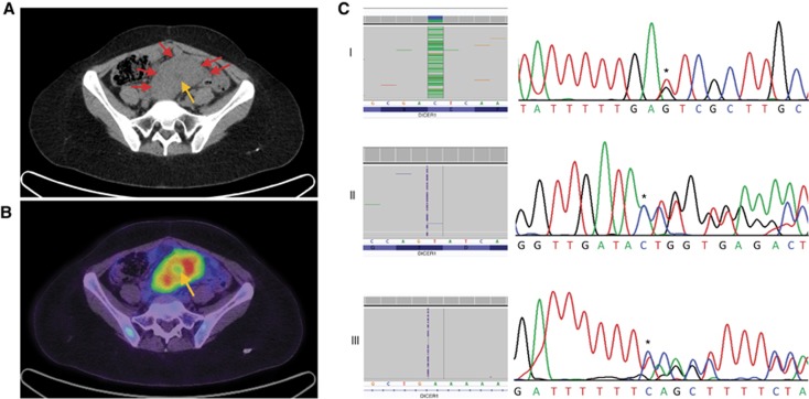
Figure 1.
Diagnostic images and mutations for case 1. (A) Axial computed tomography (CT) of pelvis demonstrating a solid pre-sacral soft tissue mass (arrows) with low attenuation signal suggesting central cystic/necrotic change (bottom right arrow). (B) Fused positron emission tomographic and CT image of pre-sacral mass demonstrating high metabolic activity as reflected by F-18 flourodeoxyglucose avidity. Central area of reduced activity coincides with area of central tumor necrosis (arrow). Following surgical resection of the tumour, the patient underwent chemotherapy (vincristine, doxorubicin, and cyclophosphamide) for 4 months and radiotherapy of the abdomen and pelvis (24 Gy). Recurrent pelvic disease was detected after an 18-month disease-free interval. Surgical resection was attempted, but complications were incurred. Three months later, recurrent disease was again noted on positron emission tomography imaging. Two cycles of irinotecan/temozolamide chemotherapy were administered. Fifty-two months after initial diagnosis, the patient succumbed to her disease. (C) The exon 25 c.5439G>T somatic mutation (Panel I), exon 11 c.1785_1786insA somatic mutation (Panel II) and intron 12 c.2040+53_2040+54insT germline mutation (Panel III) as seen in Fluidigm-derived data (left) and chromatogram (right). The mutations are indicated by an asterisk and the wild-type sequence is provided below each chromatogram. A full color version of this figure is available at the British Journal of Cancer journal online.

