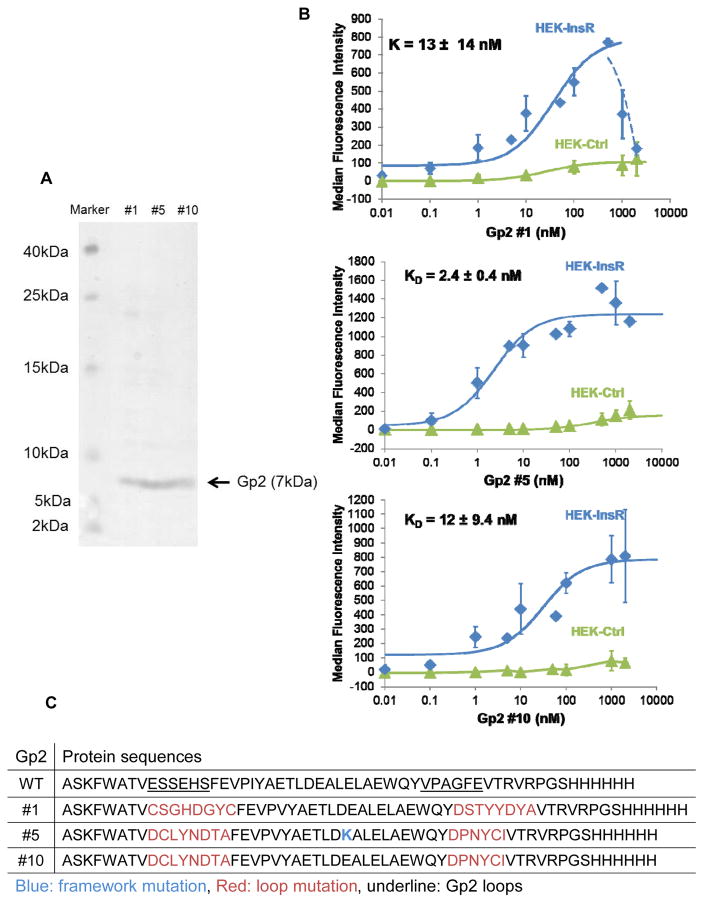
Figure 2. Affinity titration of Gp2 variants.
(A) Coomassie blue staining of soluble Gp2 clones. Identified Gp2 variants were purified using Ni-NTA resin and size exclusion filter, separated by 15% SDS-PAGE. The gel was stained with Coomassie blue. (B) HEK293T lentivirus transduced with either pLenti-InsR-GFP or pLent-GFP-ctrl were labeled with increasing concentrations of indicated soluble Gp2. Binding was detected by AF647-conjugated anti-His antibody via flow cytometry. Fluorescence signal was subtracted from the basal signal. KD values represented median ± standard deviation of three independent experiments. Some data points were not repeated and thus no standard deviation indicated. Note that HEK-Ctrl cells express modest levels of InsR (Supplementary Figure 2A), which accounts for the non-zero signal at higher concentrations. (C) Protein sequences of Gp2 variants.
 indicates a loop mutation;
 indicates a framework mutation relative to initial library.

