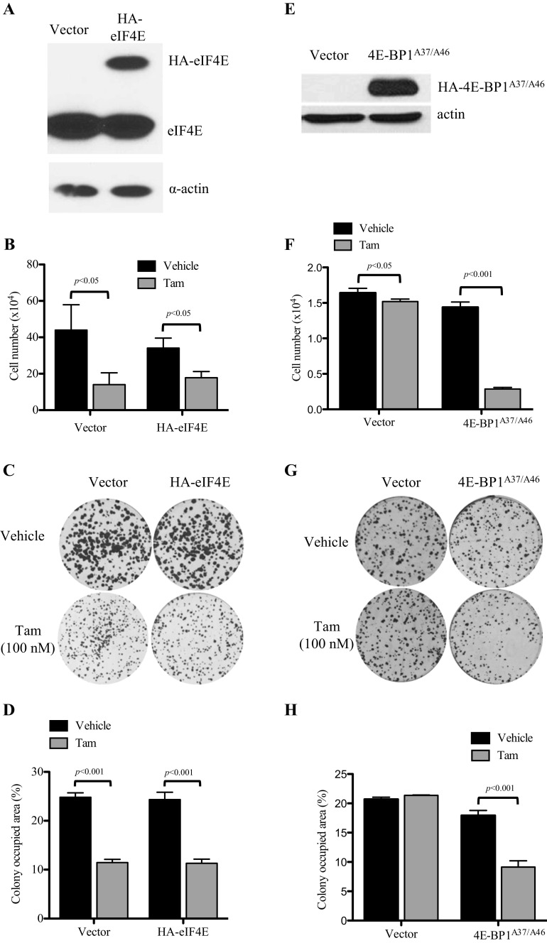
Fig. 2.
Effects of modulation of eIF4E on tamoxifen sensitivity in MCF-7L and TamR cells. a Western blot analysis demonstrating the lentivirus-mediated overexpression of HA-tagged eIF4E in tamoxifen-sensitive MCF-7L cells. Cells were transduced with a lentiviral eIF4E/GFP expression vector and flow sorted by GFP expression. b Cell growth assay to evaluate the responsiveness of MCF-7L tamoxifen-sensitive cells and eIF4E-overexpressing cells to tamoxifen. Cells were serum starved overnight before plating. Cells were grown in 5% dextran-coated charcoal (DCC)-treated FBS + treatment for 7 days and then trypsinized, stained with trypan blue, and counted. No significant difference was seen when each treatment was compared across cell lines by an unpaired t test. c Clonogenic efficiency assay examining the effect of eIF4E overexpression in response to tamoxifen in MCF-7 cells. Cells were plated and treated in full media. Colonies were allowed to grow for 10 days and then fixed and stained. Percent area was determined using GeneTools (Syngene) software and graphed in d. e Western blot showing the expression level of lentivirus-delivered HA-4E-BP1A37/A46 in TamR cells. Cells were harvested from full media and then lysed using three freeze-thaw cycles. Equal amounts of protein were resolved on an SDS-PAGE gel and immunoblotted to determine the expression of HA-4E-BP1A37/A46. f To evaluate cell growth, cells were plated in triplicate in a six-well plate and grown in 5% DCC FBS and either tamoxifen or vehicle for 5 days and then trypsinized and counted. g Stable cells were plated in triplicate in a six-well plate. After 24 h, media were replaced with media containing either tamoxifen or vehicle. Cells were allowed to grow for 10 days and then fixed and stained. Percent area was quantitated and graphed in h. A one-way ANOVA with Tukey’s post hoc test was used to compare the difference between vehicle and tamoxifen-treated groups

