
Figure 3.
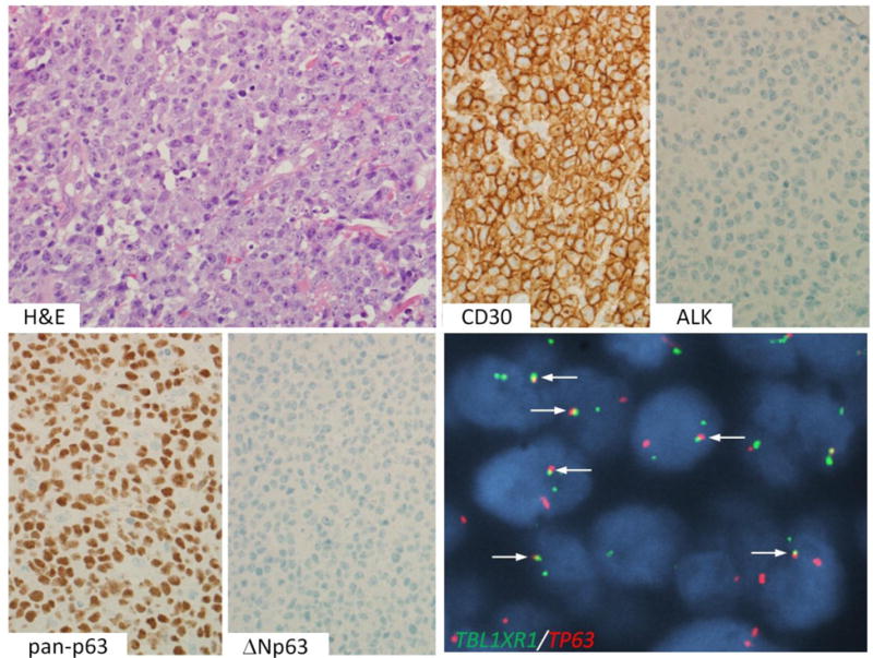
ALK-negative ALCL with TP63 rearrangement. An H&E stain shows sheets of tumor cells. By IHC, the cells are positive for CD30 and pan-p63, and negative for ALK and ΔNp63. Dual-fusion FISH shows abnormal fusion signals (arrows), indicative of TBL1XR1-TP63 fusion.