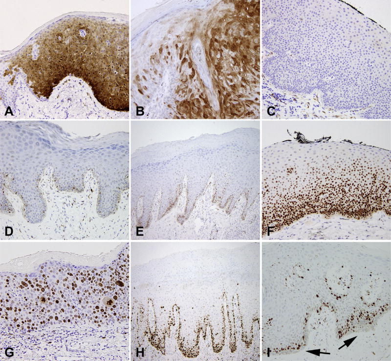
Fig. 4.
Vulvar intraepithelial neoplasia (VIN), immunohistochemical features. p16 with (A) diffuse strong block-like staining in VIN 3; (B) patchy staining in condyloma acuminatum; (C) negative in dVIN. p53 shows (D) weak patchy staining in normal skin, (E) increased basal staining in squamous cell hyperplasia, and (F) increased basal and parabasal staining in dVIN. Ki-67 shows (G) full thickness staining in VIN 3, and (H) increased basal and parabasal staining in dVIN. (I) normal skin shows no Ki-67 staining in the basal layer (arrows).

