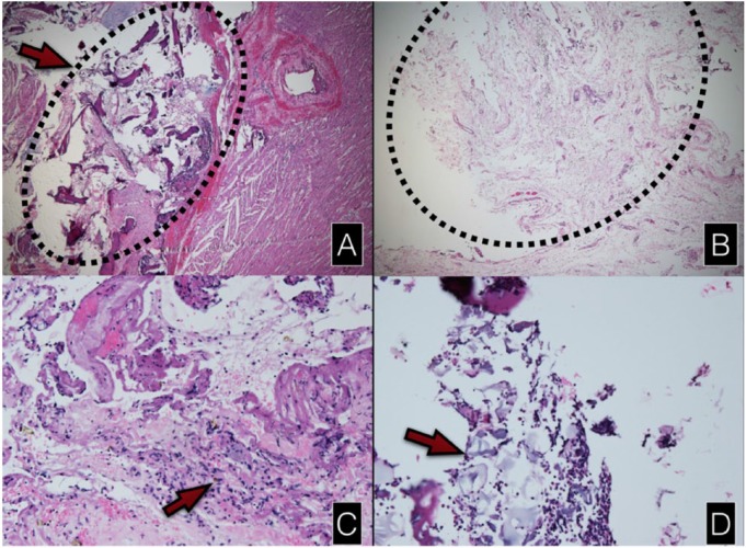
Figure 4.
Light microscopic findings. A,B: hematoxylin and eosin staining H&E, original magnification ×25. (A) Bony fragments which adhered to the epicardium in the control group. (B) The overgrowth of granulation tissue from the defect area in the collagen membrane group. (C) Giant cells demonstrated in only 1 specimen of the collagen membrane group (H&E stain, ×100). (D) The residual collagen membrane surrounded by inflammatory cells (H&E stain, ×100).

