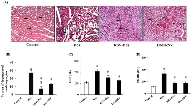
Fig 3. Resveratrol treatment rescued Dox induced myocardial damage.
(A). H & E staining: Photomicrographs of rat myocardial sections (x 200). Control group showed muscle fibers arranged in different directions (arrows), Dox group showing a wide area of widely spaced deeply acidophilic fibers including multiple disrupted (arrows), multiple thin attenuated (arrowheads) and multiple fibers exhibiting dark peripheral nuclei. RSV-Dox showed few congested blood vessels among muscle fibers, few deeply acidophilic (arrows) and few thin attenuated fibers (arrowheads) were present. In Dox-RSV group also sections showed some congested blood vessels among apparently normal muscle fibers, in addition to some deeply acidophilic fibers (arrows), some thin attenuated (arrowheads) and some fibers exhibiting dark peripheral nuclei were detected. (B). Quantitative analysis of (percent area) degenerated myocytes. (C&D). LDH and CK-MB levels in serum measured by commercial kits purchased from Stanbio Laboratory USA. Data are mean ± SD. *P < 0.05, significantly different from respective control group, #P < 0.05, significantly different from respective Dox group, $P < 0.05, significantly different from respective Dox-RSV group.

