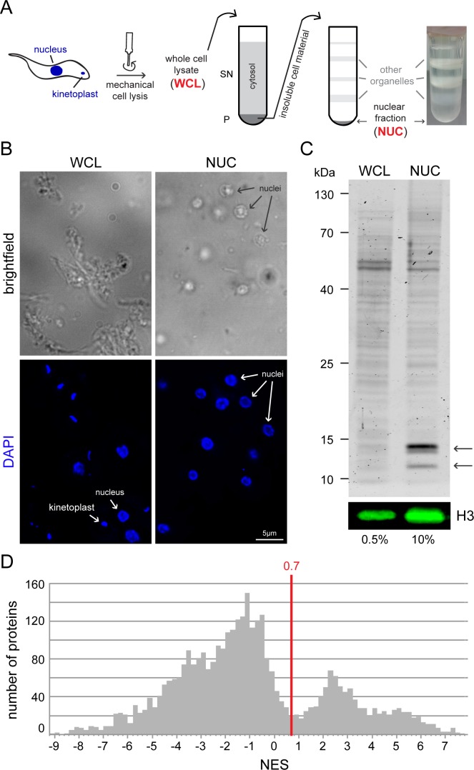
Fig 1. Purification of trypanosome nuclei.
A) Schematics of the procedure. 1•1010 procyclic trypanosome cells were mechanically lysed with a POLYTRON® homogenizer (whole cell lysate, WCL). The insoluble cell fraction which includes the nuclei was separated from the soluble fraction via a sucrose cushion and further separated on a discontinuous sucrose gradient. Various organelles and cell fragments accumulate at the interfaces of the sucrose layers and are thus separated from the nuclei, which are found in the pellet fraction (NUC). A typical picture of an ultracentrifugation tube after centrifugation is shown on the right. B) Samples of whole cell lysates (WCL) and the nuclear fraction (NUC) were stained with DAPI and microscopically analysed. In the NUC sample, isolated nuclei are clearly visible as ovoids and few other structures are present, such as remnants of flagella (brightfield image). Nuclei are intact (native shape, nucleolus is visible by absence of DAPI staining) and only few kinetoplasts are visible (DAPI image). In contrast, the WCL sample contains remnants of whole cells, including both nuclei and kinetoplasts. Note that the samples were not fixed to the slide and moved during imaging; the different channels do not completely overlap. The DAPI image is shown as deconvolved z-stack projections, the brightfield image is a single plane. C) Enrichment in histones in fraction NUC. Coomassie-stained gel loaded with 0.5% of the WCL fraction and 10% of the NUC fractions (upper panel). The arrows point to the bands corresponding to histones. In addition, histone H3 was detected by western blot (lower panel, H3). D) NES histogram: For each 0.2 NES range, the number of proteins is shown. The NES of 0.7 that was used in this work to define a nuclear protein is shown as a red line.

