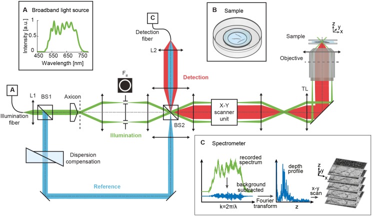
Fig 1. Schematic of the visOCM setup for C. elegans imaging.
Light from a laser source with a broad spectrum in the visible range (A, inset) is collimated by lens L1 and split by beam-splitter BS1 into a sample (green) and reference (blue) arm. In the sample arm, the axicon lens generates a Bessel-like illumination beam which is then guided to the tube lens (TL) and objective by the X-Y galvo-scanner unit. The back-reflected light (red) from the sample (B, inset) is recombined with the reference arm by beam-splitter BS2 and focused by L2 into the detection fiber. Finally, the spectrometer (C, inset), records the interference pattern which is processed to yield a depth profile of the C. elegans structure. The data processing steps are illustrated in S1 Fig. Scale bars: 25 μm.

