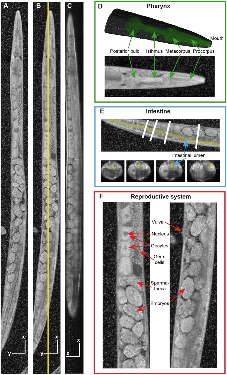
Fig 3. Anatomy of the C. elegans as revealed by visOCM.
(A, B) En face projections at two different depths, and (C) side view at the location highlighted in (B). Scale bars indicate 50 μm. (D) Top: A 3D rendered model of the head with the pharynx highlighted in green. Bottom: Maximum-intensity projection through the entire animal’s head. (E) En face view (top) and corresponding transverse sections (bottom), with the lumen of the intestine highlighted in yellow. (F) Zoom regions of the reproductive system showing germ cells, oocytes, spermatheca, embryos and the vulva. The 3D sub-micrometer resolution and the intrinsic contrast of our technique enable a clear and detailed visualization of tissue structures down to the sub-cellular level (see also S2 Video).

