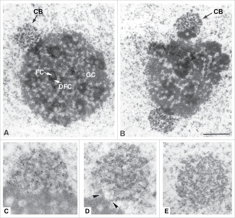
Figure 1.

Electron Micrographs showing the relationship between nucleoli and Cajal bodies in rat trigeminal ganglion neurons. A and B) CBs juxtaposed to nucleoli. Note the typical structure of the nucleolus with fibrillar centers (FC), dense fibrillar components (DFC) and granular components (GC). In B, the CB (arrow) appears to be associated with a segregated mass of dense material (asterisk) in close proximity to the nucleolar surface C to D) Immunoelectron localization of coilin. With the anticoilin antibody a high density of immuno-gold particles (small black dots) specifically decorated the dense coiled threads of CBs. In C, an extensive portion of the CB is directly apposed on the dense fibrillar component of the nucleolus, whereas the CB of D maintains a minimal connection with this nucleolar component (arrowheads), and the CB of E appears free into the nucleoplasm. Adapted with permission from.4
