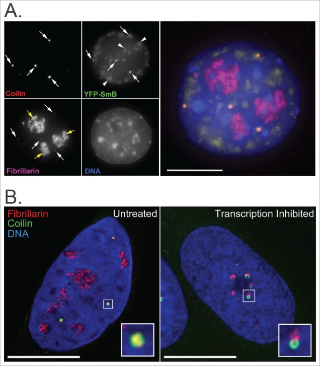
Figure 2.

Immunofluorescence images showing co-localization of nucleolar and CB factors. (A) Fibrillarin and SmB co-localize with Coilin at CBs (white arrows) in mouse embryonic fibroblast cells. Coilin (red) was detected using anti-coilin primary and anti-rabbit-Cy5 secondary antibodies, Fibrillarin (magenta) using anti-Fib primary and anti-mouse-TRITC secondary antibodies, SmB (green) by overexpression of YFP-tagged SmB for 48 hours and DNA (blue) by DAPI staining. Fibrillarin is also present in nucleoli (yellow arrows). SmB is also present in speckles (arrowheads). Bar = 10 μm. (B) In untreated U2OS cells, a subset of Fibrillarin (red) accumulates with Coilin (green) in CBs (boxed region, expanded as inset). Upon inhibition of Pol I- and Pol II-mediated RNA transcription by Actinomycin D treatment (2.5 ug/ml for 2 hrs), Fibrillarin and Coilin aggregate in distinct peri-nucleolar caps (boxed region, expanded as inset). Fibrillarin was detected using anti-fibrillarin primary and anti-mouse-Alexa568 secondary antibodies, Coilin using anti-coilin primary and anti-rabbit-Alexa488 secondary antibodies and DNA by DAPI staining. Bar = 10 μm.
