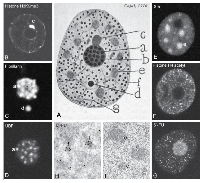
Figure 1.
Cajal's original drawing of the neuronal nucleus. (A) The scheme shows the nuclear structures identified in pyramidal neurons from the cerebral cortex (“Original drawing at the Cajal Institute, C.S.I.C., Madrid”). Panels B-I illustrate the equivalent nuclear components identified at the present time in mammalian neurons. (B) Nucleolus-attached heterochromatin (“Levi basophilic grume, c”). (C) nucleolus and CB (“argyrophilic nucleolar spherules,” a;” “ground substance, b” “accessory body, d”). (D) Fibrillar centers of the nucleolus (“argyrophilic nucleolar spherules, a”). (E) Nuclear speckles (“hyaline grumes, e”). (F) Nuclear microfoci of active chromatin immunolabeled for the acetylated histone H4 (“neutrophil grains, f”). (G) Nuclear foci of 5′-fluorouridine (5′-FU) incorporation into nascent RNA (“neutrophil grains, f”). (H) Ultrastructural characterization of nuclear foci of 5′-FU incorporation (I) Fine structure of interchromatin granule clusters in a neuron (“hyaline grumes e”). Note that Cajal's drawing shows the nucleus enclosed by 2 parallel lines. (C and E from Lafarga et al., Chromosoma 2009; G from Casafont et al., Acta Neuropathol 2011, reproduced with permission from © Springer).3,99

