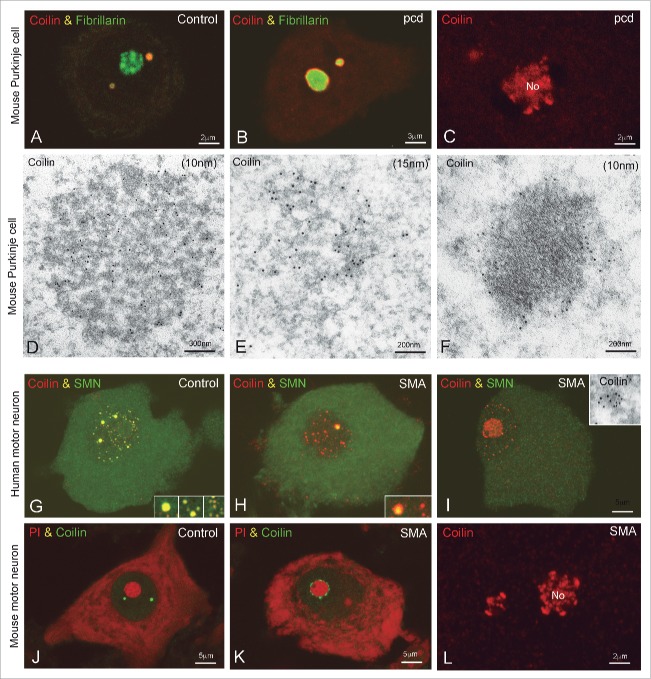
Figure 7.
Reorganization of Cajal bodies in Purkinje cells of the pcd mice and motor neurons from SMA. (A-C) Confocal microscopy images from squash preparations of Purkinje cells from wild type (A) and pcd mice (B, C) co-stained for fibrillarin and coilin (A, B) or single immunostained for coilin (C). (A) Fibrillarin and coilin colocalize in a CB from a control Purkinje cell. (B, C) At advanced stages of Purkinje cell degeneration, with nucleolar segregation and fragmentation, coilin appears redistributed as a thin ring surrounding the segregated masses of fibrillarin or inside the nucleolus (No). (D) In wild type Purkinje cells, CBs show the typical morphology of coiled threads immunogold labeled for coilin. (E) CB with an irregular morphology and loosely arranged threads in a degenerating Purkinje cell. (F) Gold particles of coilin immunoreactivity are also observed surrounding an electrondense mass presumably corresponding to a segregated portion of the dense fibrillar component of the nucleolus. (G-I) Double immunostained for coilin and SMN on dissociated motor neurons from control and SMA samples. (G) In a control neuron coilin concentrates in several large CBs and numerous mini-CBs where it colocalizes with SMN (inset). (H) This motor neuron from an SMA patient shows a large CB immunolabeled for coilin and SMN and numerous coilin-positive and SMN-negative mini-CBs (inset). (I) SMA motor neuron with an eccentric nucleus, intranucleolar accumulation of coilin and numerous coilin microfoci free of SMN. (inset) Detail of a coilin microfocus with immunogold electron microscopy. (J-L) Motor neurons from a wild type mouse (J) and an SMA mouse model (K and L). (J, K) Co-staining for coilin and propidium iodide (PtdIns) illustrates the typical organization of CBs in the control neuron (J) and the redistribution of coilin as perinucleolar caps in the SMA motor neuron (K). (L) Intranucleolar localization of coilin (No) in an SMA motor neuron. (A; B, D-F, from Baltanas et al., Brain Pathol 2011, reproduced with permission from © John Wiley and Sons; G-I from Tapia et al. Histochem Cell Biol 2012, reproduced with permission from © Springer.60,96

