Figure 3.

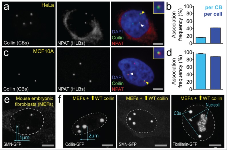
Cajal bodies are dynamic structures. (a-d) HeLa cells and human breast epithelial cell line MCF10A cultured for 3 d and fixed using 4% paraformaldehyde were stained with antibodies to detect CBs (CB marker protein coilin, green) and HLBs (histone transcription factor NPAT, far red) and imaged using the Opera 5020 high-content automated confocal microscopy system (PerkinElmer, Waltham MA). Briefly, nuclear segmentation and automated spot detection was performed using proprietary PerkinElmer Acapella software. CB-HLB distances were exported and a threshold (4 pixels between spot centers) was applied to quantify CB-HLB associations. N = 2, approx. Five,000 cells. Associations were normalized on a per-CB and per-cell basis. (e) Maximal intensity projection image of mouse embryonic fibroblasts (MEFs) expressing endogenous WT coilin transfected with plasmids containing SMN-GFP. CB size was measured using Zeiss Zen 2 software. (f) Maximal intensity projection images of MEFs transfected with plasmids containing WT (untagged) mouse coilin plus SMN-GFP, mouse coilin-GFP or fibrillarin-GFP (to co-stain both CBs and nucleoli). CB size was measured using Zeiss Zen 2 software.
