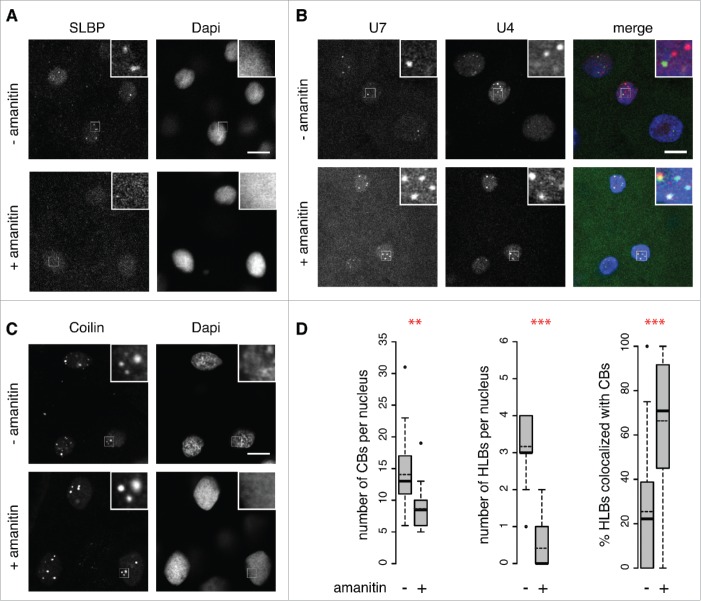
Figure 3.
Separation of CBs and HLBs is transcription-dependent. Zygotic transcription was blocked by injection of α-amanitin into 1-cell embryos. Single Z-stacks of confocal series from fixed blastula stage embryos. Untreated (- amanitin) and treated (+ amanitin) embryos are labeled with antibodies specific for SLBP (A) or coilin (C); co-injection of U4 (red) and U7 snRNAs (green) provides an independent view of CBs and HLBs, respectively (B). White square indicates area shown magnified in each inset (4x magnification). Scale bars equal 10 μm. (D) Quantification of CBs per nucleus (coilin immunostaining, as in C; data from n=18 embryos from 3 independent experiments at 4hpf), HLBs per nucleus (SLBP, as in A; data from n = 18 untreated and n = 17 α-amanitin injected embryos from 3 independent experiments at 4hpf), and of the overlapping signals between HLBs and CBs (U4 and U7 snRNAs, as in B; n = 31 nuclei from untreated embryos and n = 16 nuclei from α-amanitin injected embryos at 3hpf). Distribution, mean number (dashed line) and median number (bold line) are shown. Asterisks indicate statistical significance (Welch 2-sample t-test, P = 0.004702 for CBs, P = 1.043e-10 for HLBs and P = 7.208e-05 for U4/U7 snRNA).

