
Figure 4.
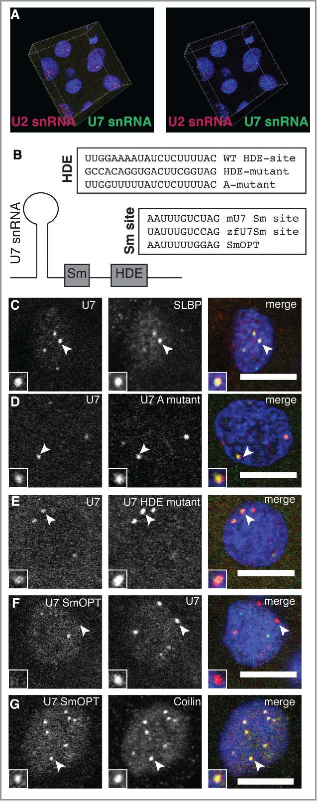
Sm ring composition specifies snRNA localization to CBs and HLBs. (A) Co-injection of U2 (magenta) and U7 (green) snRNAs shows independence of CBs and HLBs in 3 dimensions. Projection of the fluorescent data collected by confocal imaging is shown in the left panel; rendering of CB and HLB centers of mass, obtained through the use of Imaris analysis software, is shown in the right panel. (B) Scheme for testing the contribution of base-pairing to histone transcripts through mutational analysis of the U7 snRNA HDE site is shown. To test the contribution of the Sm-ring, the specialized Sm site of mouse U7 (mU7) was mutated to Sm-OPT, previously shown to target the spliceosomal Sm ring to U7 snRNA.47 (C-G) Confocal sections showing the localization patterns of wild-type U7 snRNA compared to other markers and mutants, as indicated. Scale bars equal 10 μm. Arrowheads point to foci magnified 2x in the insets.