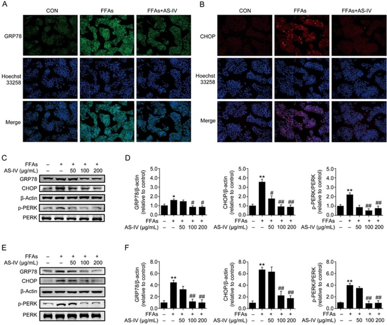
Figure 5.
AS-IV alleviates FFA-induced ER stress in hepatocytes. HepG2 cells and primary murine hepatocytes exposed to FFAs (1 mmol/L) were treated with or without AS-IV (50–200 μg/mL) for 24 h. (A) Representative images of immunofluorescence staining for the ER stress marker GRP78 at 200× magnification in HepG2 cells. (B) Representative images of immunofluorescence staining for the ER stress marker CHOP at 200× magnification in HepG2 cells. (C) Representative immunoblots for GRP78, CHOP, Thr981p-PERK and PERK in HepG2 cells. β-Actin was used as an internal control. (D) Densitometric analyses of the band intensity ratios for GRP78/β-actin, CHOP/β-actin and Thr981p-PERK/PERK in HepG2 cells. (E) Representative immunoblots for GRP78, CHOP, Thr981p-PERK and PERK in primary murine hepatocytes. β-Actin was used as an internal control. (F) Densitometric analyses of the band intensity ratios for GRP78/β-actin, CHOP/β-actin and Thr981p-PERK/PERK in primary murine hepatocytes. Data are presented as the mean±SEM of three independent experiments (n=3). *P<0.05, **P<0.01 compared with the control group; #P<0.05, ##P<0.01 compared with the FFA group. FFAs, free fatty acids; AS-IV, astragaloside IV.

