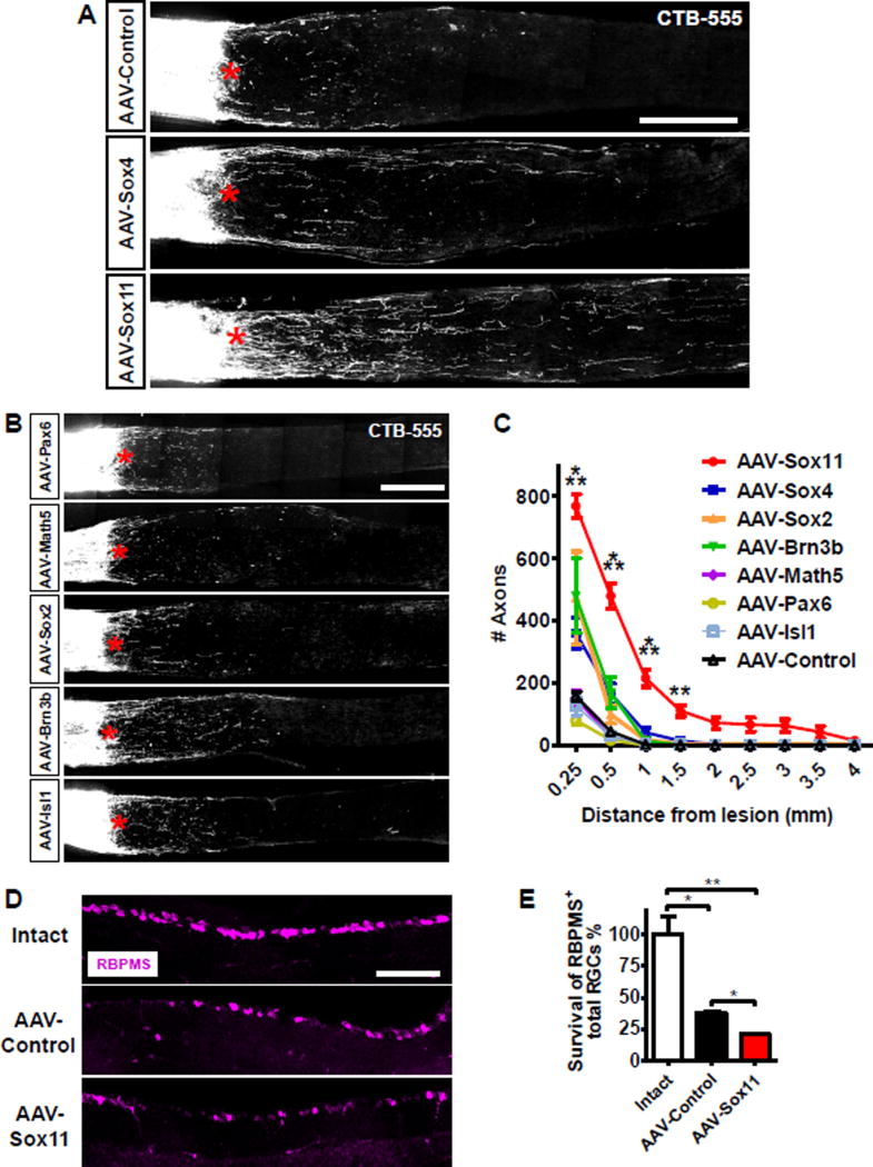
Figure 1. Over-expression of Sox11 promotes optic nerve regeneration.
(A, B) Representative images of optic nerve sections showing CTB-labeled axons in wild type mice with intravitreal injections of AAV expressing PLAP, Sox4, and Sox11 (A), and Pax6, Math5, Sox2, Brn3b, and Isl1 (B) at 2 weeks after optic nerve injury. The crush site is indicated with a red asterisk. Scale bars in (A) and (B) represent 250 µm.
(C) Quantification of regenerating axons from A and B. Data are expressed as mean ± SEM (n = 3–12). *** P < 0.001 (ANOVA with Bonferroni posttests; relative to AAV-Control).
(D) Representative retinal sections stained with anti-RBPMS antibodies from intact retina, or the retina at 2 weeks after injury with prior injection of AAVs expressing PLAP (Control) or Sox11. Scale bar represents 100 µm. (E) Quantification as in D. RBPMS-positive cells in the ganglion cell layer were imaged and quantified for two sections per retina, and normalized to length counted. Data are expressed as mean ± SD (n = 3–4). * P < 0.05, ** P < 0.01 (ANOVA with Bonferroni posttests).

