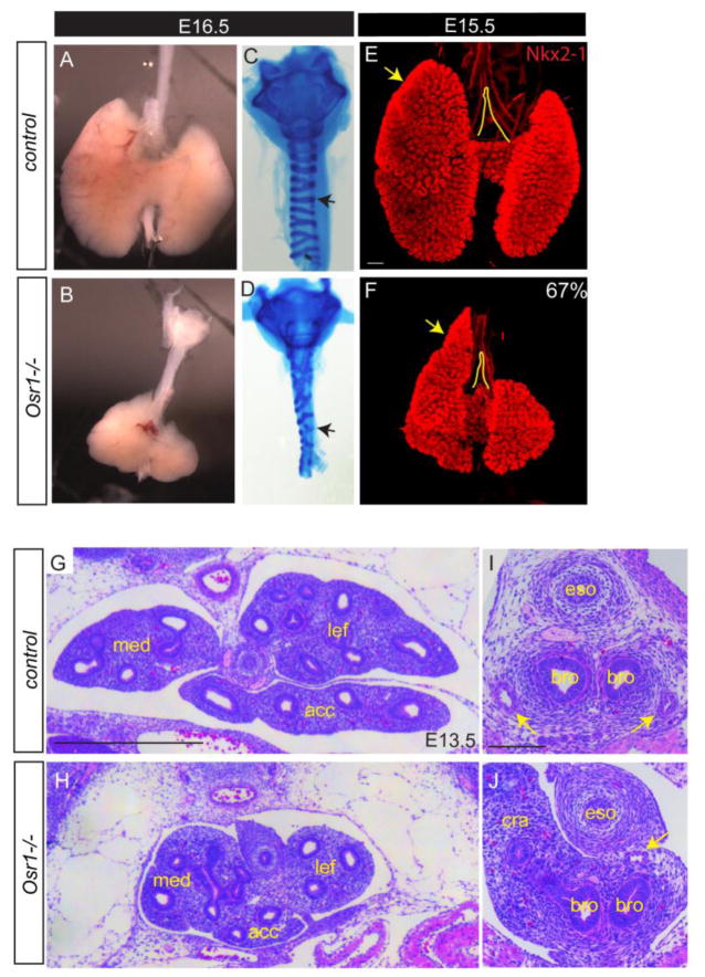
Fig. 2.
Osr1 mutants display lung hypoplasia, dysmorphyc lobulation and mesenchymal differentiation defects. (A–B) Bright field view of dissected esophagus, trachea and lung. (C–D) Alcian blue staining of dissected trachea. Arrows show broken and disorganized cartilage rings in Osr1 mutant compared to control. (E–F) Whole mount IF of Nkx2-1 in wildtype and mutant lungs. Arrows point to the anterior extension of the cranial lobe relative to the trachea bifurcation. Scale bar: 200 μm. (G–J) H&E staining of transverse sections lung (G–H, scale bar: 500 μm) and main bronchi (I–J, scale bar: 100 μm) regions. Arrows indicate pulmonary arteries. Med, medial lobe; lef, left lobe; acc, accessory lobe; eso, esophagus; bro, bronchus; cra, cranial lobe.

