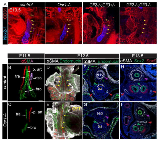
Fig. 4.
Mesenchymal development including pulmonary arteries and smooth muscle differentiation in foregut is dependent on Osr1. (A) Whole mount IF staining in E10.5 embryos. Yellow arrows mark the vascular plexus connecting the out flow tract to the lungs. Scale bar: 50 μm. (B–C) αSMA whole mount staining of dissected foregut. Epithelial staining (not shown) is used to guide the identification of different smooth muscle cell structures. Different regions of the staining are isolated and pseudo-colored using Imaris. (D–E) Whole mount staining of the dissected foregut with the heart attached. Arrows point to the pulmonary arteries. Scale bars: 100 μm. (F–I) IF on transverse sections at the trachea level (F–G) and the main bronchi level (H–I). Purple arrows indicate esophagus SMC, green arrows indicate tracheal SMC, while red arrows indicate pulmonary arteries SMC. OFT, out flow tract; p. art, pulmonary artery; tra, trachea; eso, esophagus; bro, bronchi. Scale bars: 50 μm.

