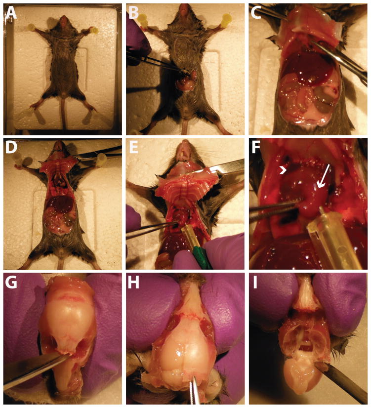
Figure 2.
Mouse necropsy and tissue collection. A) The mouse is pinned down to the surgical board in all four paws. B) An incision is made into the ventral abdomen. C) The incision is extended up to the diaphragm, which is carefully cut open. D) The incision is extended on both sides to open the chest cavity. Hemostats are used to hold the cavity open. E) A needle is placed into the mouse’s left ventricle for perfusion of saline and paraformaldehyde. F) Close-up image of heart at perfusion. Arrowhead indicates the mouse’s right atrium, which is cut at the start of the perfusion. Arrow indicates the left ventricle, where the needle is placed for perfusion. G) After removing the mouse’s head and exposing the skull, a small incision is made through the skull between the eye sockets. H) A cut is made up the midline of the skull, starting at the cervical wound, and the cut skull is peeled off using forceps. I) After full removal of the dorsal skull, the brain is gently removed using a spatula.

