Figure 1.
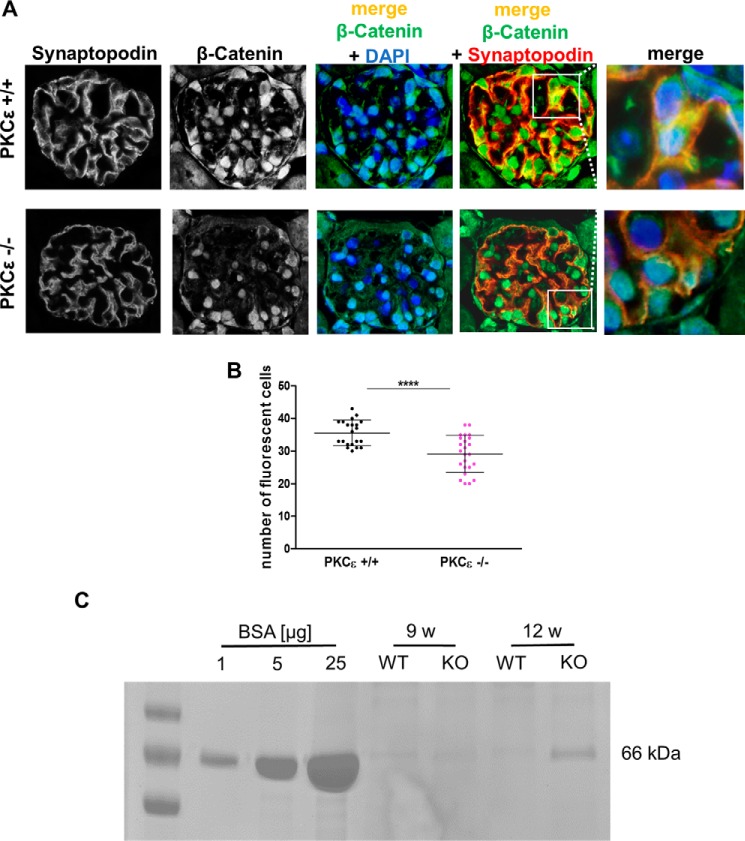
β-Catenin expression in glomeruli of wild-type and PKCϵ knockout mice. A, immunofluorescence staining of murine glomeruli at 12 weeks of age in PKCϵ+/+ and PKCϵ−/− mice. Co-staining with β-catenin (green), the podocyte marker synaptopodin (red), and DAPI (blue) shows expression and localization within glomeruli. B, semiquantitative analysis of the number of cells with β-catenin expression in PKCϵ+/+ and PKCϵ−/− mice (****, p < 0.0001). C, SDS-PAGE/Coomassie gel staining of urine from wild-type and PKCϵ knockout mice at 9 and 12 weeks (w). BSA at 1, 5, and 10 μg/ml served both as a control and standard. Data are mean ± S.D. of at least three different independent experiments.

