Figure 4.

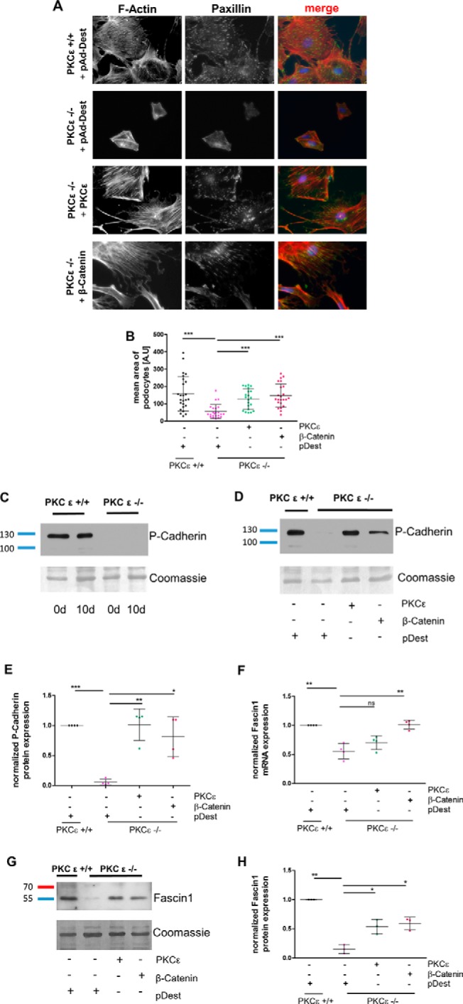
Overexpression of β-catenin rescues the phenotype of PKCϵ knockout podocytes. A, murine PKCϵ−/− podocytes were transduced with adenoviral vectors expressing either pAd-Dest, PKCϵ, or β-catenin. Immunofluorescence staining of the actin cytoskeleton with phalloidin and against paxillin was performed. B, the cell size of phalloidin-marked podocytes was assessed in ImageJ for determination of the mean area (A.U, area unit). C, Western blot analysis of P-cadherin expression in murine podocytes from day (d) 0 and day 10. D, PKCϵ−/− podocytes were transduced with adenoviral vectors expressing either pAd-Dest, PKCϵ, or β-catenin and then analyzed for P-cadherin expression with Western blot analysis. E, quantification of P-cadherin band intensity using total protein analysis with Coomassie staining of the membrane. F, quantitative RT-PCR of RNA extracted from cells of the same experiment as in A was used to assess fascin1 mRNA expression and normalized to HPRT. G, Western blot analysis of the same experiment as in A for fascin1 protein expression. H, quantification of band intensity by total protein analysis with Coomassie staining (*, p < 0.05; **, p < 0.05; ***, p < 0.0001; ns, not significant; Student's t test). Data are represented as mean ± S.D. of at least three different independent experiments.
