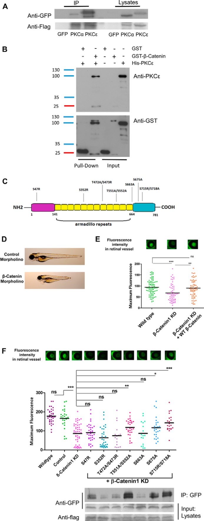
Figure 5.

PKCϵ is a functional binding partner of β-catenin, and the interaction is indispensable for filtration barrier function. A, FLAG immunoprecipitation (IP) of HEK cells transduced with a FLAG-tagged β-catenin construct and either a GFP, GFP-tagged PKCα (positive control), or GFP-tagged PKCϵ construct. Immunoblotting against FLAG shows successful transfection. B, in vitro direct interaction between β-catenin and PKCϵ was examined in a GST pulldown experiment with pure recombinant GST-β-catenin and His-PKCϵ. His-PKCϵ was incubated with either GST or GST-β-catenin. The pulldown fractions were analyzed by Western immunoblotting against PKCϵ and GST. C, schematic illustrating β-catenin protein structure, which is divided into the N-terminal, Armadillo repeats, and C-terminal parts. The suggested phosphorylation sites of PKCϵ are shown. D, individual larvae at 120 hpf, indicating control morpholino–injected fish with no edema and β-catenin morpholino–injected fish with edema and a dorsalized phenotype. E, murine β-catenin-mutants were tested in the zebrafish model by injecting the capped mRNA into fertilized zebrafish eggs at one- to four-cell–stage embryos. The transgenic zebrafish produce a vitamin D-binding protein fused with GFP that, under normal conditions, accumulates in the retina and is quantified 120 hpf by measuring the fluorescence level. Reduced fluorescence indicates a disturbed glomerular filtration barrier. The graph shows GFP fluorescence intensity of the eye assay of wild-type, β-catenin1 morpholino and co-injection of β-catenin1 morpholino with a human wild-type RNA construct. F, group mean fluorescence intensities of the eye assay. A comparison of the combined injection of β-catenin morpholino plus β-catenin-mutant results was conducted against β-catenin morpholino injection alone (*, p < 0.05; **, p < 0.01; ***, p < 0.001; ns, not significant). Binding of each β-catenin mutant to GFP-tagged PKCϵ was tested in vitro with FLAG immunoprecipitation in transfected HEK cells. Immunoblotting was performed against GFP and FLAG.
