Figure 6.
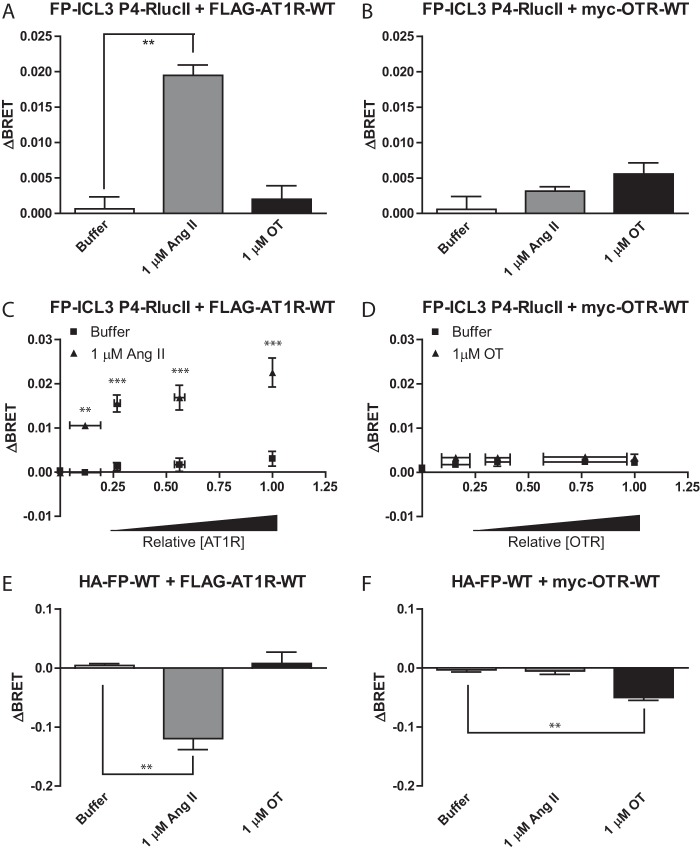
Activation of Gαq by the oxytocin receptor does not induce a similar conformational change in FP. A and B, HEK 293 cells were transfected with FP-ICL3 P4-RLucII and AT1R-WT (A) or OTR-WT (B). Cells were stimulated with the indicated ligand/concentration and the change in BRET (ΔBRET) due to ligand stimulation is reported. C and D, HEK 293 cells were transfected with a constant amount of the cDNA for FP-ICL3 P4-RLucII and increasing amounts of AT1R-WT (C) or OTR-WT (D) cDNA. Cells were stimulated with 1 μm Ang II (C) or oxytocin (OT) (D) and the change in BRET (ΔBRET) due to ligand stimulation is reported. Ligand-induced changes in BRET plotted against the relative AT1R (C) or OTR (D) surface expression as assessed by in-cell western blots normalized to the highest expression level. E and F, HEK 293 cells were transfected with a BRET-based Gαq activation sensor, FP-WT, and AT1R-WT (E) or OTR (F). Cells were stimulated with the indicated ligand/concentration and the change in BRET (ΔBRET) due to ligand stimulation is reported. A, B, E, and F, bars represent the mean of 3 independent biological replicates and error bars represent S.E. Dunnett's test was performed comparing to buffer where, ** = p < 0.01. C and D, a two-way analysis of variance was performed, n = 3. For C, both factors of relative expression and stimulation as well as the interaction were significant. For D, both factors of relative expression and stimulation were significant but the interaction was not. Bonferroni corrected t-tests were performed to compare the buffer versus Ang II or OT at each relative expression level, where ** = p < 0.01 and *** = p < 0.001.

