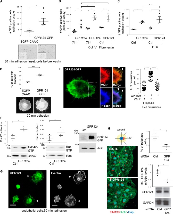
Figure 1.
GPR124 promotes cell adhesion, polarity, filopodia formation, and activation of Cdc42 and Rac1 GTPases. A, GPR124-dependent cell adhesion. Adhesion assays were performed using serum-starved COS7 cells expressing GFP-tagged GPR124 or EGFP–CAAX, used as control. Cells were fixed with 4% PFA, and images were taken with a Nikon Eclipse Ti inverted microscope and analyzed with NIS-Elements software. The graph shows the mean ± S.E. (error bars) of adherent GFP-positive cells for each condition (Student's t test; *, p < 0.05; n = 4). Representative fields below the graph show adherent cells at 30 min, and the inset shows all EGFP-positive cells before non-adherent cells were washed away. B, GPR124 shows an additive effect on cell adhesion to collagen IV and fibronectin. COS7 cells transfected with EGFP–CAAX (control plasmid (Ctrl)) or GPR124–GFP were plated on plastic, collagen IV, or fibronectin for 30 min. Adherent cells were analyzed using ImageJ software. The graph shows the mean ± S.E. of adherent GFP-positive cells (*, p < 0.05; ***, p < 0.001). Statistics were performed using one-way ANOVA followed by Tukey's multiple-comparison post hoc test (n = 3). C, inhibition of Gi heterotrimeric proteins does not alter GPR124-dependent cell adhesion. Control (Ctrl; EGFP–CAAX) or GPR124–GFP–transfected COS7 cells were treated overnight and during 30 min adhesion assays with vehicle or pertussis toxin (PTX; 200 ng/μl). Fixed adherent cells were analyzed using ImageJ software. The graph shows the mean ± S.E. of GFP-positive cells and was analyzed by one-way ANOVA followed by Tukey's multiple-comparison post hoc test (*, p < 0.05; n = 3). D, GPR124 induces filopodia formation. The morphology of COS7 cells expressing GPR124–GFP was monitored in cells left adhering for 30 min and compared with control COS7 cells expressing EGFP–CAAX. Statistics were performed by Student's t test (***, p < 0.0005; n = 3). Representative cells are shown at the bottom of the graph. E, GPR124 localizes at VASP-positive filopodia in adhering cells. COS7 cells transfected with GPR124–GFP and mCherry–VASP, a filopodial marker, were subjected to adhere for 30 min, fixed, stained for F-actin, and analyzed by confocal microscopy. About 75% of VASP-positive filopodia protrusions per cell were also positive for GPR124–GFP (graph in the right panel). A representative cell is shown at the left. Cellular protrusions positive for either GPR124 and VASP or GPR124 and F-actin are indicated by arrowheads in the merged images. At least 30 cells were analyzed by confocal Leica TCS SP8 using a ×100 objective. The graph shows the mean ± S.E. (n = 3). One-way ANOVA followed by Tukey's multiple-comparison post hoc test was performed for statistics (****, p < 0.0001). F, GPR124 promotes activation of Cdc42 and Rac GTPases. Serum-starved cells expressing either GPR124–GFP or control EGFP–CAAX cells were lysed and incubated with PAK-N beads to capture active Cdc42 and Rac1. Cdc42-GTP and Rac1-GTP were identified by Western blotting. Cdc42-GTP was increased in COS7 cells expressing GPR124. The graph shows the mean ± S.E. of normalized Cdc42 and Rac activation (Student's t test; *, p < 0.05; n = 3). G, GPR124 is localized at cell projections of adhering endothelial cells. Endogenous GPR124 (top) was localized with filamentous actin (bottom) at cell projections of HUVECs (white arrowheads) left to adhere for 30 min (similar results were observed in 36 of 60 cells). Bar, 20 μm. H, GPR124 is required to polarize HUVECs. HUVECs with siRNA knockdown of GPR124 (siGPR124) were less polarized at the edge of the wound than siRNA control (siCTL). Nuclei were stained with DAPI (blue), and filamentous actin was visualized with green fluorescent phalloidin. Cells in which GM130 (a Golgi marker; red) did not localize within a 120° angle facing the wound were considered as non-polarized cells. At least 100 cells near the wound edge were randomly examined. The percentage of polarized cells is shown in the top graph (right). Data are represented as mean ± S.E. (Student's t test; *, p < 0.05; n = 3). GPR124 knockdown was confirmed by quantitative RT-PCR (right, bottom graph).

