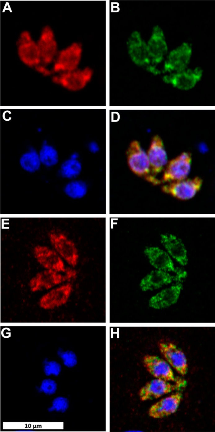
Figure 6.

Toxoplasma stained for ectopically expressed TgSPT1-TY (A, AlexaFluor594, red) and endogenous TgSPT1 (E, AlexaFluor594, red); ectopically expressed ER marker GFP-HDEL (B and F, AlexaFluor488, green); and DNA (C and G, DAPI, blue). Co-localization of TgSPT1, ectopically expressed and endogenous, with GFP-HDEL is shown in merge of A and B (D, yellow) and E and F (H, yellow), respectively. The scale bar is equivalent to 10 μm.
