Figure 3.
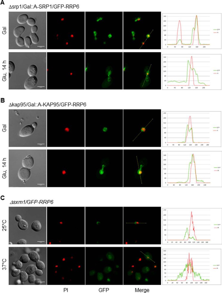
Inhibition of karyopherins expression affects the subcellular localization of GFP-Rrp6. A, laser-scanning confocal microscope images show the subcellular localization of GFP-Rrp6 after inhibition of Srp1 expression for 14 h in glucose medium in Δsrp1/GAL::SRP1 cells. Analysis of GFP-Rrp6 relative to PI using ImageJ is shown on the right. Green lines represent GFP, and red lines represent PI. Localization of GFP in Δsrp1/GAL::SRP1 cells is shown in supplemental Fig. S2. B, analysis of the subcellular localization of GFP-Rrp6 after inhibition of Kap95 expression for 14 h in glucose medium in Δkap95/GAL::KAP95 cells. Analysis of GFP-Rrp6 relative to PI using ImageJ is shown on the right. C, analysis of the subcellular localization of GFP-Rrp6 in Δsxm1 cells growing at 25 or 37 °C. Because GFP-Rrp6 expression is lower in this strain growing at 37 °C, gain of the confocal microscope had to be increased for the Rrp6 signal to be detected. Importantly, this signal was above background levels. Scale bars, 4 μm.

