Figure 4.
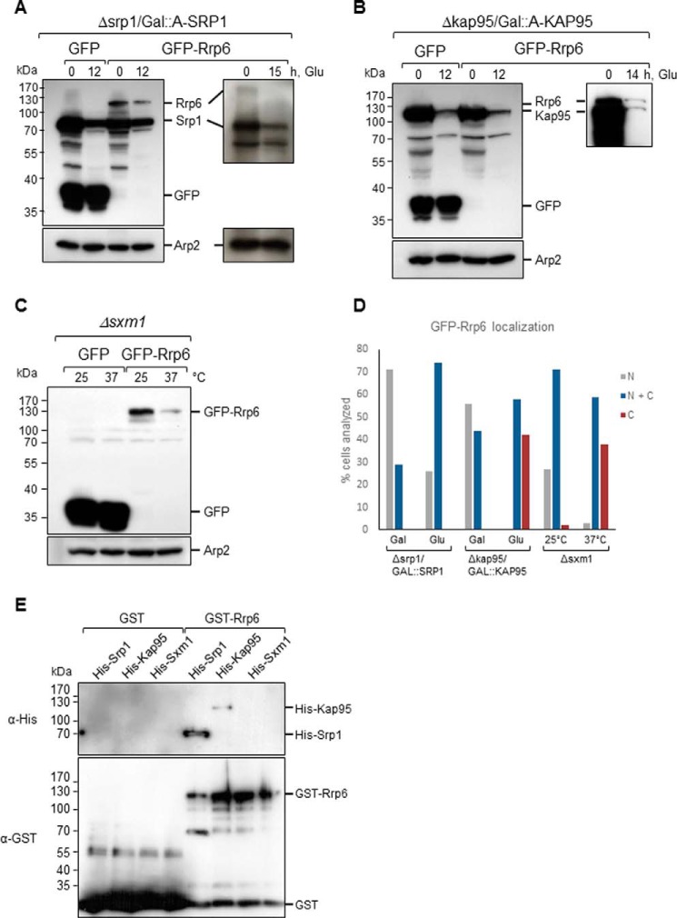
Inhibition of karyopherins expression affects the levels of GFP-Rrp6. Western blotting of total cell extract from karyopherin mutants expressing either GFP or GFP-Rrp6 growing in galactose (Gal)- or glucose (Glu)-containing medium was performed with antibody against GFP, which also allowed the detection of ProtA-Srp1 and ProtA-Kap95. A, Δsrp1/GAL::A-SRP1 growing in glucose shows the lower levels of ProtA-Srp1 after 12 or 15 h in glucose. GFP-Rrp6 also decreases upon inhibition of Srp1 expression. B, Western blot of total cell extract from Δkap95/GAL::A-KAP95 expressing either GFP or GFP-Rrp6 shows the lower levels of ProtA-Kap95 in glucose. GFP-Rrp6 and ProtA-Kap95 have very similar molecular masses. A longer run and exposure for separation and visualization of the bands are shown on the right-hand side. C, Western blot of total cell extract from Δsxm1 expressing either GFP or GFP-Rrp6 growing at 25 or 37 °C shows that the expression levels of GFP-Rrp6 are lower at 37 °C. Western blotting with antibody against Arp2 was used as an internal control. D, quantitative analysis of cells expressing GFP-Rrp6. Approximately 100 cells of each strain were analyzed by fluorescence microscopy, and cells that showed protein localized to the nucleus (N), present both in nucleus and cytoplasm (N + C), or visible mainly in the cytoplasm (C) were counted. Numbers correspond to the percentage of cells showing each phenotype. E, pulldown of Srp1 and Kap95 with Rrp6. GST or GST-Rrp6 bound to glutathione-Sepharose beads was incubated with His-Srp1- or His-Kap95-containing extracts. Elution fractions are shown. The same membrane was incubated with antibody against His tag and subsequently with antibody against GST tag. The figure shown is representative of three independent experiments.

