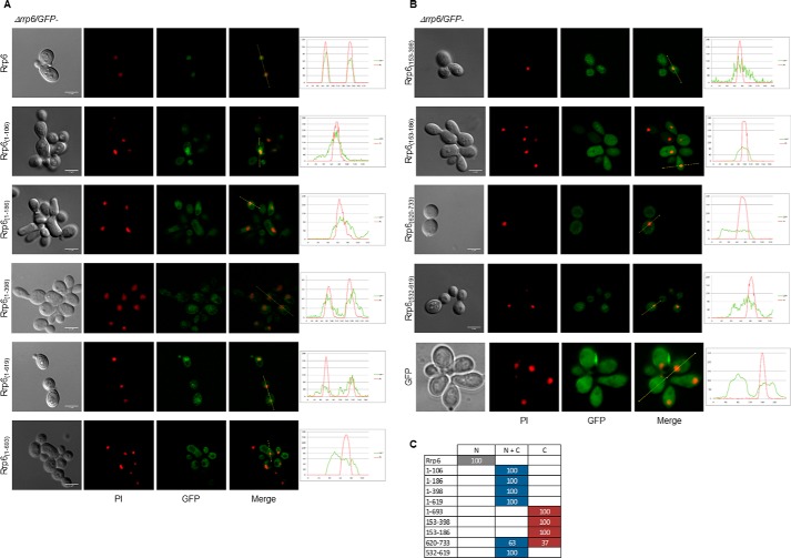
Figure 7.
Presence of canonical NLS is not the only determinant of Rrp6 nuclear import. Laser-scanning confocal microscope images show the subcellular localization of the Rrp6 deletion mutants expressed in Δrrp6 cells growing at 25 °C. A, GFP-Rrp6, Rrp6(1–106), Rrp6(1–186), Rrp6(1–398), Rrp6(1–619), and Rrp6(1–693). B, Rrp6(153–398), Rrp6(153–186), Rrp6(620–733), Rrp6(532–619), and GFP. Various mutants accumulate in the nucleus although to different levels. Quantification is shown on the right. C, quantitative analysis of cells expressing GFP-fused Rrp6 mutants. N, protein concentrated in the nucleus; N + C, protein present both in nucleus and cytoplasm; C, protein visible mainly in the cytoplasm. Numbers correspond to the percentage of cells showing each phenotype. Scale bars, 4 μm.

